
Ana Maria Mihalcea, MD, PhD - May 11, 2024 ∙ Paid ∙ Source
Likes: 98 | Comments: 60 | Reposts: 24 | ALL OTHER POSTS
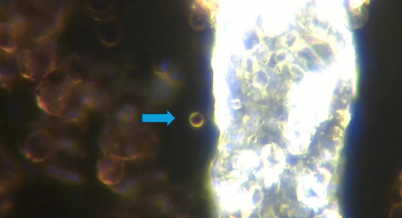
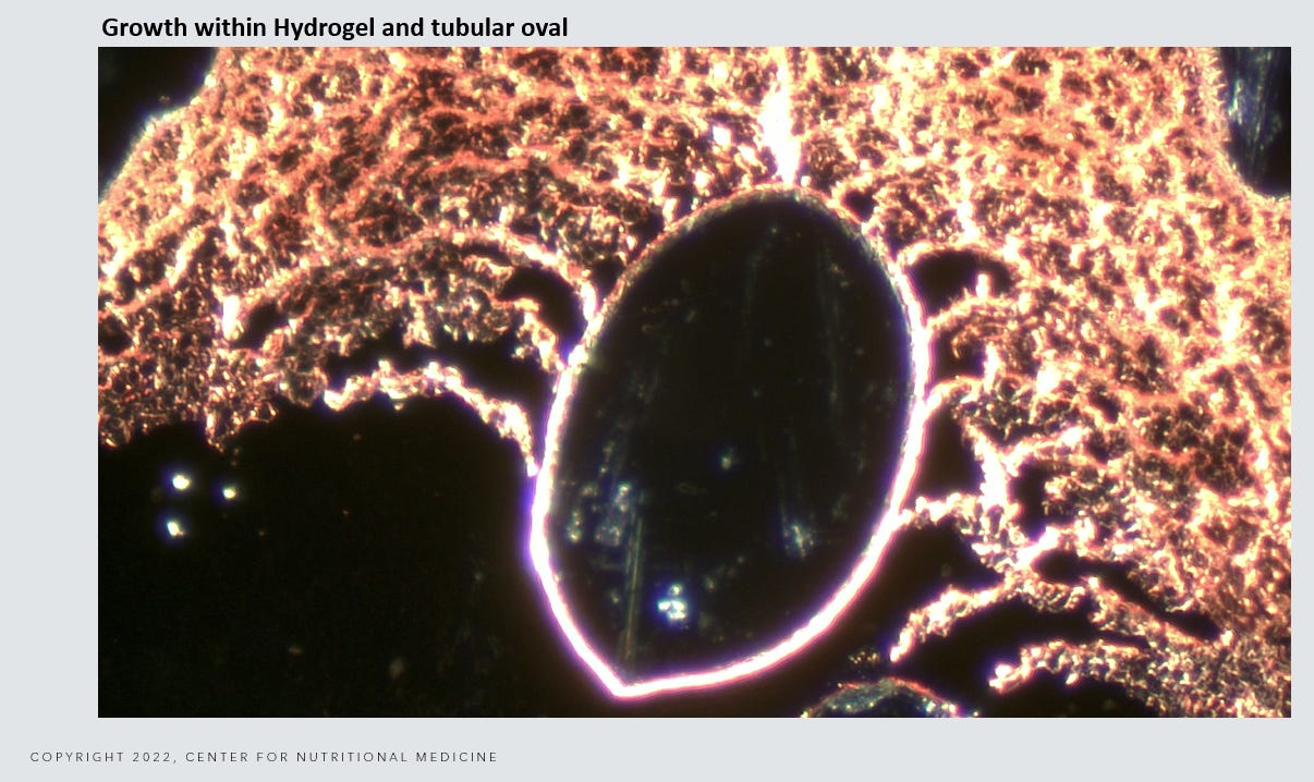
Image: Darkfield Live Blood Analysis - Large vesicle containing many micellar structures of different sizes.
In this post, I wanted to review the scientific literature of transition states from tubular micelles to spherical micelles to large vesicles. Some people think if you see that the vesicle is dissolving in the blood or on the microscopy slide that the blood is cleaned. That is not necessarily so. The vesicle simply has changed states back to a micellar or tubular state. The self assembly nanotechnology or drug delivery system that delivers a cargo payload can assemble and disassemble when certain external conditions are met. Lets review the literature on this:
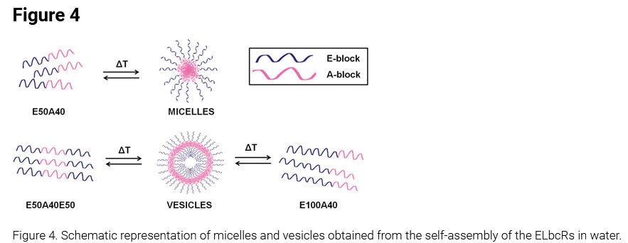
The possibility of obtaining different self-assembled nanostructures in reversible systems based on elastin-like block corecombinamers is explored in this work. The results obtained show how an evolution from a more common micellar structure to a hollow vesicle can be attained simply by changing the block arrangements and lengths, even when other molecular properties, such as molecular weight or mean polarity, remain essentially unchanged. This work sheds light on the possibility of obtaining hollow nano-objects, based on elastin-like recombinamers, which can assemble and disassemble in response to a change in their surroundings. This kind of system can be an example of how high precision in the genetic production of synthetic macro-molecules can be used, on an exclusive basis, to control the shape and size of their derived nano-objects.
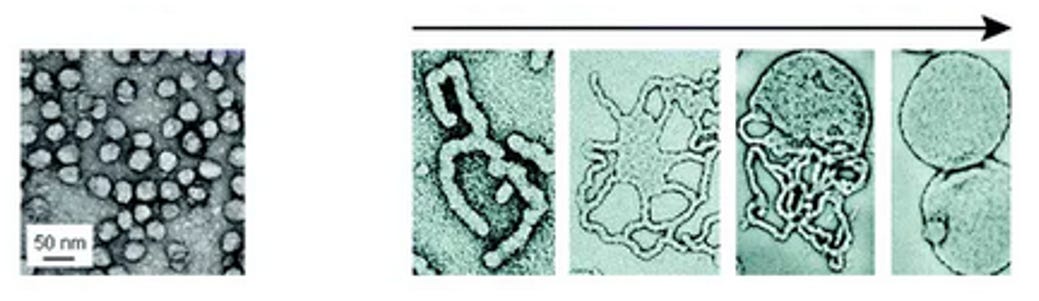
Here is a figure based on temperature differential. Filaments can grow into micelles or into vesicles and the process is reversible, but that does not mean the nanotechnology is gone. When the external conditions change again, it can morph again:
The control of the transformation can be chemically induced as well - the process is externally controllable:
However, although micelles are most easily obtained by way of many different molecular interactions, other more complex shapes often require the existence of precise molecular interactions that drive the self-assembled objects to adopt hollow vesicular or tubular structures. (1-3) Typically, such interactions are of the hydrogen-bonding or ion-pair type and give rise to strong and stable cores in which the molecular segments involved show characteristics such as crystallinity, swelling, directionality, and rigidity, among others. (4) However, although such relatively strong interactions can be tuned to model the shape of the self-assembled object, they tend to lead to practically irreversible self-assembly.
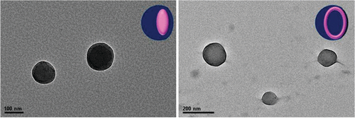
You can see that from the denser micelles hollow vesicles can grow ( polymersomes).
In Darkfield Live Blood it looks like this spherical object. This is not an artificial cell, it is a vesicular transport for a payload cargo.
Remember that polyacrylamide was one of the stealth nanoparticles in the Moderna patent. You can make micellar structures that change form from many different polymer chemicals. The article discusses that the conversion of micelles to vesicles can take minutes or weeks depending on what chemicals or external tuning control mechanisms are used. The micelles serve as building blocks that fuse with other structures and can build solid objects. All processes are changeable, however that again does not mean the problem is gone, it just changed forms:
Does PNIPAM block really retard the micelle-to-vesicle transition of its copolymer?
The well-known coil-to-globule transition of poly( N -isopropyl acrylamide) (PNIPAM) at its LCST lasts as short as hundred of seconds with fully reversibility. However, for the PNIPAM-containing block copolymers , thermal transformation from micelles to vesicles caused by the conformation transition of PNIPAM took as long as several weeks, even at the temperatures much higher than the LCST, and without satisfactory reversibility. In the literature, this slow process has been attributed to the strong interchain hydrogen bonding in PNIPAM, which retards the transition. In this work, asymmetrically modified PNIPAM (Mw 10K), i.e . C 12 -PNIPAM-CA with a hydrophobic hydrocarbon chain –C 12 H 25 ( C 12 ) at one end and a hydrophilic carboxyl group –COOH ( CA ) at the other, was prepared and found to form micelles with a core of the lightly associated hydrocarbon chains. When temperature is increased to the LCST of PNIPAM, t he transformation from micelles to vesicles can be realized within 30 min, while the reverse process only takes a few minutes. Based on full monitoring of the transition process, it is proposed that the micelles serve as building blocks in constructing the vesicles via processes of combination, fusion, and etc ., in which only local conformation adjustment of PNIPAM is involved.
Here you can see a figure of this transition and you can see the vesicle is filled with a cargo:
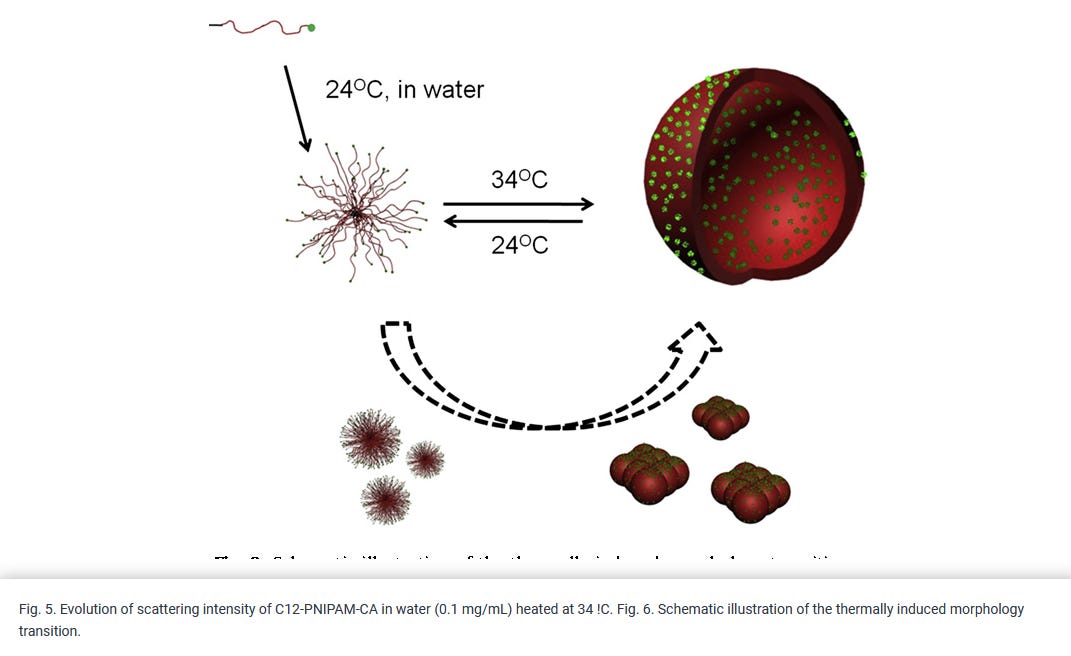
This is what you can see here, a large vesicle with a cargo of smaller micelles and nanobots that are self assembling the large polymer structure:
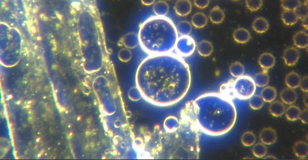
Image: C19 unvaccinated Darkfield Live blood self assembly process
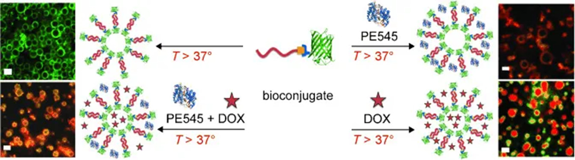
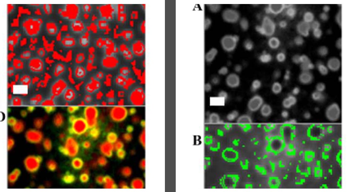
These larger vesicles and micelles can change form - and are used for drug delivery systems but can also be used for biosensing technology. Here you see images of many polymerosomes used in synthetic biology. Compare the images with the picture up top from the Darkfield live blood microscopy.
Polymersomes provide a good platform for targeted drug delivery and the creation of complex (bio)catalytically active systems for research in synthetic biology. To realize these applications requires both spatial control over the encapsulation components in these polymersomes and a means to report where the components are in the polymersomes. To address these twin challenges, we synthesized the protein–polymer bioconjugate PNIPAM- b -amilFP497 composed of thermoresponsive poly( N -isopropylacrylamide) (PNIPAM) and a green-fluorescent protein variant (amilFP497). Above 37 °C, this bioconjugate forms polymersomes that can (co-)encapsulate the fluorescent drug doxorubicin and the fluorescent light-harvesting protein phycoerythrin 545 (PE545).
PISA: construction of self-organized and self-assembled functional vesicular structures
Within this membrane a state far-from-equilibrium is maintained, which provides energetic conditions enabling complex functionalities such as adaptation and reproduction through internal metabolic processes. Polymerization-induced self-assembly (PISA) is a rapidly-developing method to produce polymersomes, or polymer vesicles, which can be considered as a synthetic analogy to vesicles with a lipid bilayer. Though to express the range of functions that systems that exhibit the basic functionalities of early life is indeed a challenge, PISA offers a particularly robust “boot up” mechanism which solves some of the challenges associated with the emergence of life. Recent advances in the exploitable chemistries applied to PISA expand the scope of complex nano-to-micron scale vesicular structures autonomously generated from a homogeneous mixture in mild conditions. This could ultimately lead to the achievement of synthesizing objects capable of mimicking basic behaviors and functions of natural life.
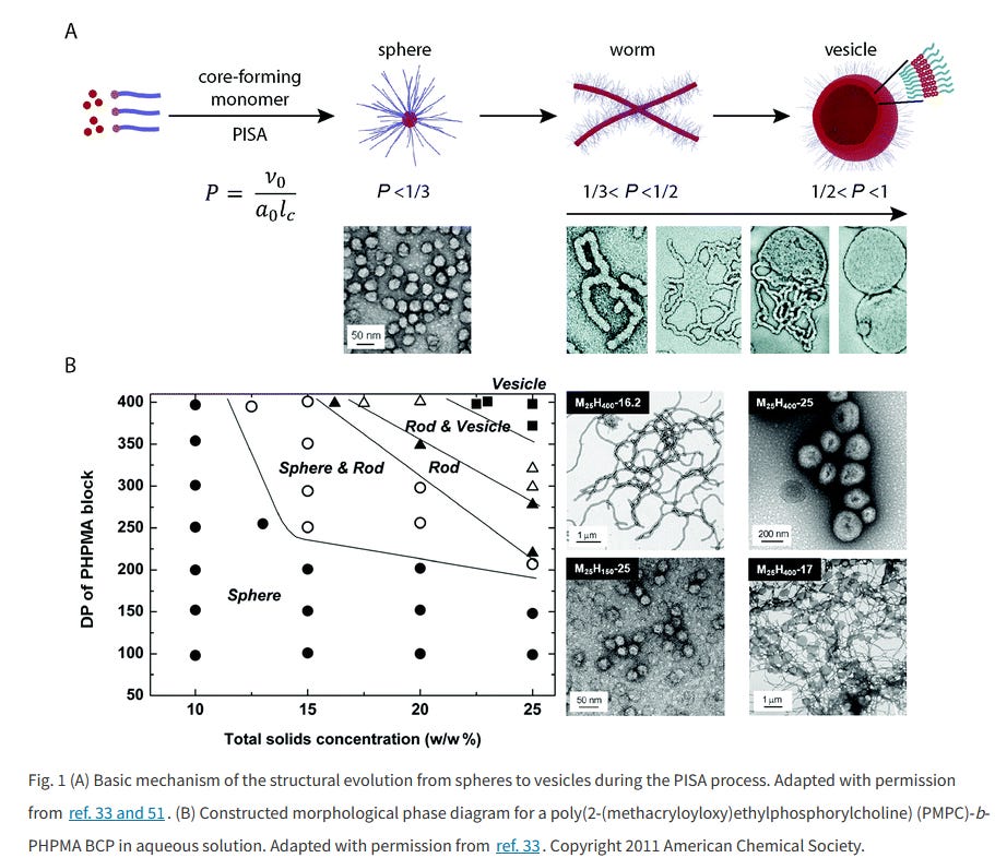
Here you can again see the transitions from spheres to “worms” ( also called tubular micelles, I call them filaments), into larger vesicles.
Lets compare this to what we see in the live blood.
You can see this process here in one of the slides from Dr. Sandy Corlett:
The transition can also be induced by chemicals:
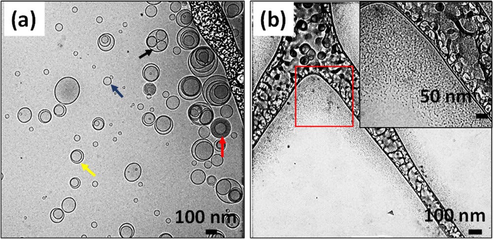
Figure 4. Nanostructures formed by pure lecithin and pure Tween 80. (a) Cryo-TEM of lecithin, with the blue arrow pointing to a unilamellar vesicle, yellow arrow pointing to a bilamellar vesicle, red arrow pointing to a multilamellar vesicle, and black arrow pointing to an oligo-vesicular vesicle. (b) Cryo-TEM of Tween 80 micelles. Amphiphile mixtures are prepared at an overall amphiphile (lecithin or Tween 80) concentration of 2.25 wt %.
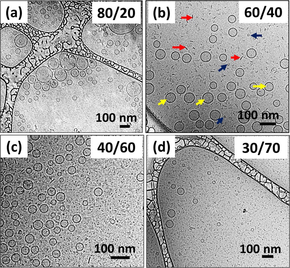
Figure 5. Cryo-TEM images of the samples at (a) 80/20, (b) 60/40, (c) 40/60, and (d) 30/70 L/T weight ratios. Large unilamellar vesicles are formed at 80/20. Bicelles and vesicles coexist at 60/40, 40/60, and 30/70 L/T weight ratios. The red, blue, and yellow arrows point to bicelles on an edge-on orientation and bicelles on a face-on orientation and vesicles, respectively. Mixtures are prepared at an overall amphiphile (lecithin + Tween 80) concentration of 2.25 wt %.
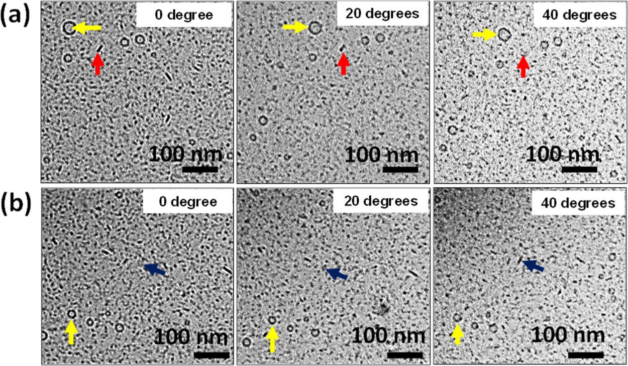
Figure 6. Change in nanostructures’ morphology with tilting cryo-TEM stage. (a) Change in the bicelle morphology from edge-on orientation to face-on orientation (red arrows). (b) Change in the bicelle morphology from face-on orientation to edge-on orientation (blue arrows). The vesicle morphology (yellow arrows) stays the same (spherical symmetry). LT mixture (30/70) prepared at an overall amphiphile (lecithin + Tween 80) concentration of 2.25 wt %.
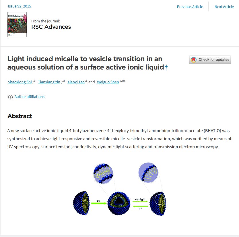
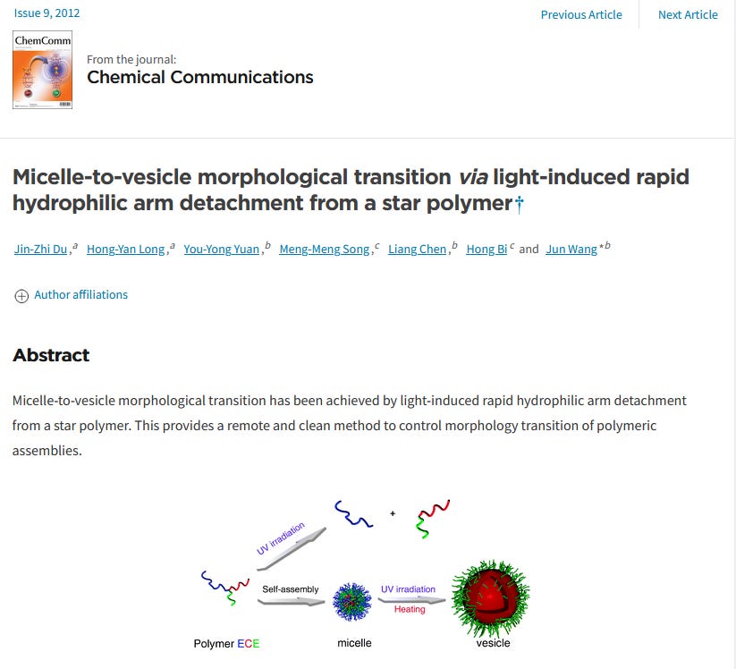
You can use light to do the same:
Here you can see another chemical modality of dissolving the larger vesicles. Again, that does not mean the technology is gone, it just has changed states:
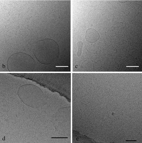
Fig. 1. Solubilization of DOPC (2.5 mM) liposomes by Triton X-100 and decylmaltoside as function of the detergent concentration (a). The turbidity change after increase of the Triton X-100 concentration (0.8 mM/step) is almost instantaneous, whereas the turbidity change after decylmaltoside addition (1 mM/step) (arrows) is slow between R sat and R sol (inset a). At 4.8 and 6.2 mM detergent (filled symbols) samples were taken for cryo-electron microscopy (b–e). Solubilization with Triton X-100, open bilayer fragments and spherical mixed micelles coexists (b), solubilization with decylmaltoside, closed vesicles and worm-like micelles coexists (c). After 1 week equilibration the Triton X-100 solubilized sample did not change. (d) In the decylmaltoside sample the vesicles disappeared and only worm-like micelles remained. Bar 100 nm.
You can do the same with pH. In alkaline blood you will see less of the large structures that I call construction sites. This is why in people on an alkaline diet the blood quality and contamination is certainly better than in an acidic environment.
Colloidal assemblies, for example, micelles,1-3 vesicles,4,5 or fibers,6 that are responsive to changes in surrounding conditions such as pH, temperature, or UV light could be potentially used as templates for materials synthesis7-9 or drug carriers and delivery devices. 1 0-12 Of particular interest are pH-sensitive vesicles, whose closed bilayer structure allows for the encapsulation and targeted release of molecules.
Summary:
I wanted to explain this process of transition states, assembly and disassembly, and morphing of what we see in the blood based on environmental factors. It does not mean the blood is clean or necessarily that there was success in dissolving the technology. You can change the states of this technology through modifying the local environment, but again it does not indicate that the technology is gone. If you let the blood cool and keep the slide, the polymerization process will be accelerated. The cause is the temperature change, which is why I do not recommend using EBOO or methods that would cool the blood as that can accelerate the polymerization process of nanoscale technology that was not seen in the process of the procedure. The clots that practitioners see in the tubing coagulate due to the temperature change, which is what embalmers like Richard Hirschman also observed - that some of the rubbery clots formed after death. That makes sense, because with the level of blood contamination we see now, we should have a lot more deaths. When I have drawn blood from patients in a 30 cc syringe and left it to cool overnight, that is when the rubbery clot developed as a function of time, meaning the polymerization process was accelerated exponentially by the temperature drop. This is consistent with the literature in polymer chemistry.
Regardless the methods, it is always great news if after treatments there is an improvement in the blood quality and contamination.
At this time, if I see one nano or microrobot in the blood sample, I know the individual is contaminated, because that one bot that I see came from the nanoscale that is self replicating and it will construct larger polymer networks. Of course, one drop is not an absolute representative of 6 liters of blood, but it does give us some form of evaluating levels of self assembly nanotechnology contamination.
Likes: 98 | Comments: 60 | Reposts: 24 | Share Options: Copy Link
TreeTomato - May 11
Arlene’s Newsletter
Given that these entities respond to Wi-Fi, etc., they respond to frequencies. in that particular frequencies or waveforms will interact with the entities.
Given that each body organ has a frequency of its own and that each material vibrates at its own frequency would it be possible to direct a frequency at a particular entity (sphere, filament, etc.) that affects only that entity (if each entity is individually addressable via some encoded ID)? If so, then it should be possible to direct the frequencies of potent substances at the entity without affecting the body. These might include strong acids that would otherwise destroy human tissue.
Just spitballing in my amateur armchair.
REPLY | 5 replies
PaulieG - May 11
Ana, a Ted Talk from 2013 on the nanotech, describing the various structures you and others are finding, and their purposes. Very helpful for identification
https://m.youtube.com/watch?v=EGgbeqNFqqo
https://m.youtube.com/watch?v=EGgbeqNFqqo
REPLY | 0 replies
58 more comments...
anaunited anapost