Ana Maria Mihalcea, MD, PhD - Apr 02, 2024 ∙ Paid ∙ Source
Likes: 114 | Comments: 50 | Reposts: 21 | ALL OTHER POSTS
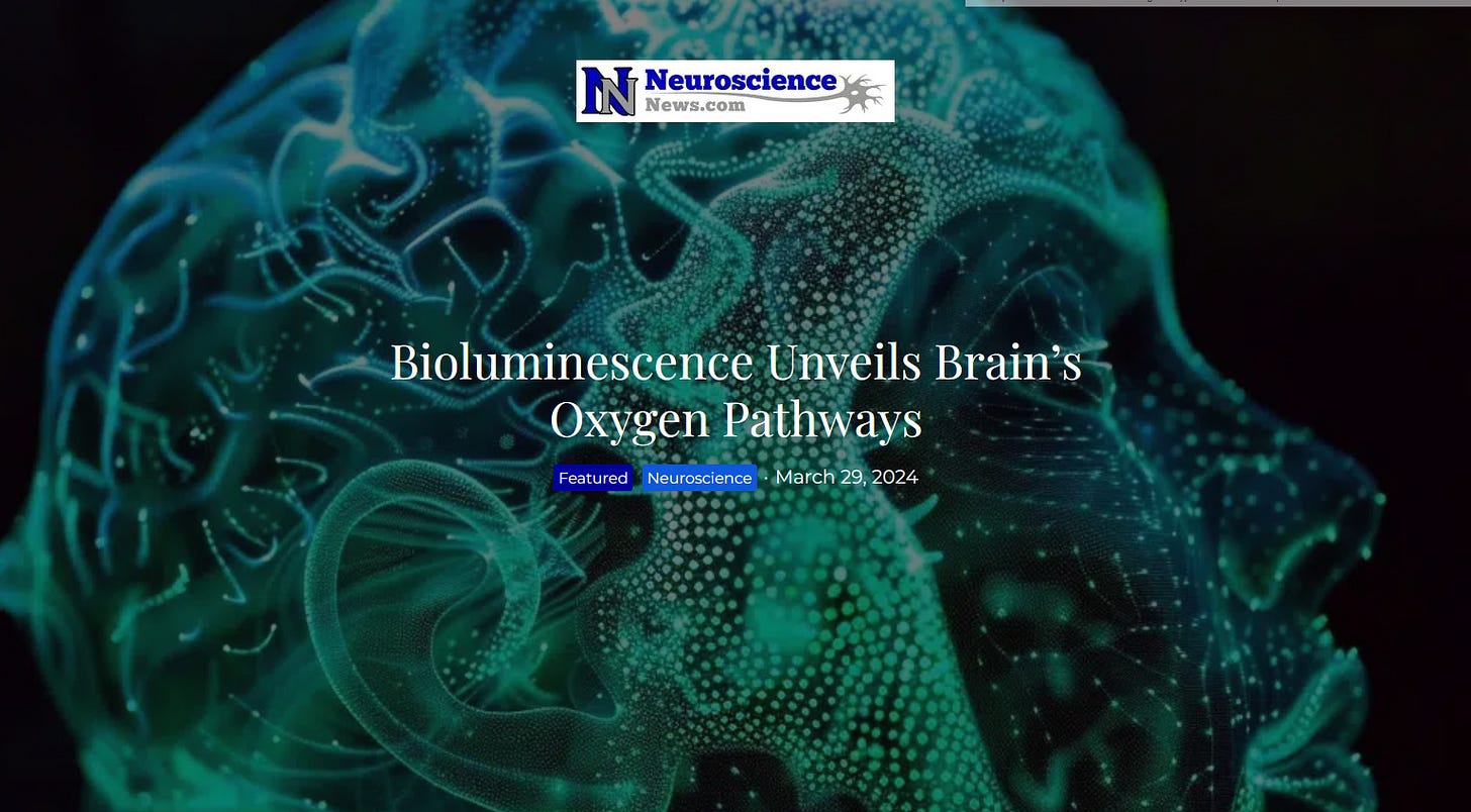
Bioluminescence Unveils Brain’s Oxygen Pathways
You might remember my recent posts on the fluorescent skin findings of the C19 injected and less so in the C19 un- injected due to shedding.
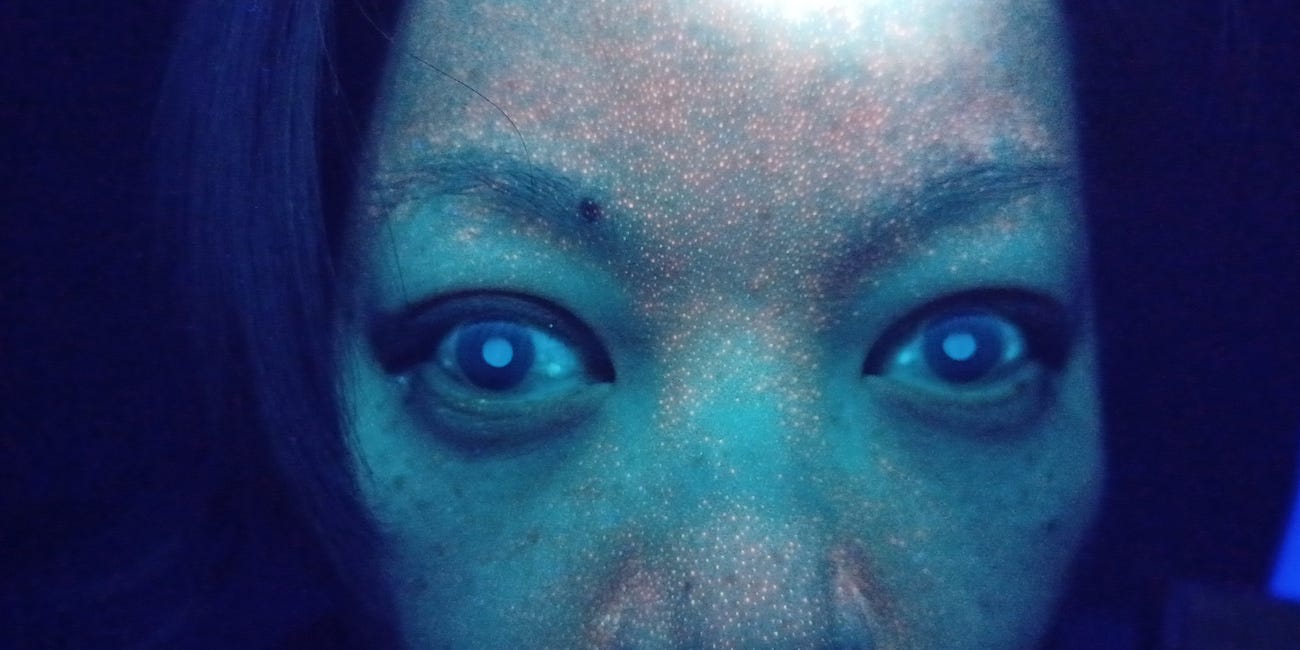
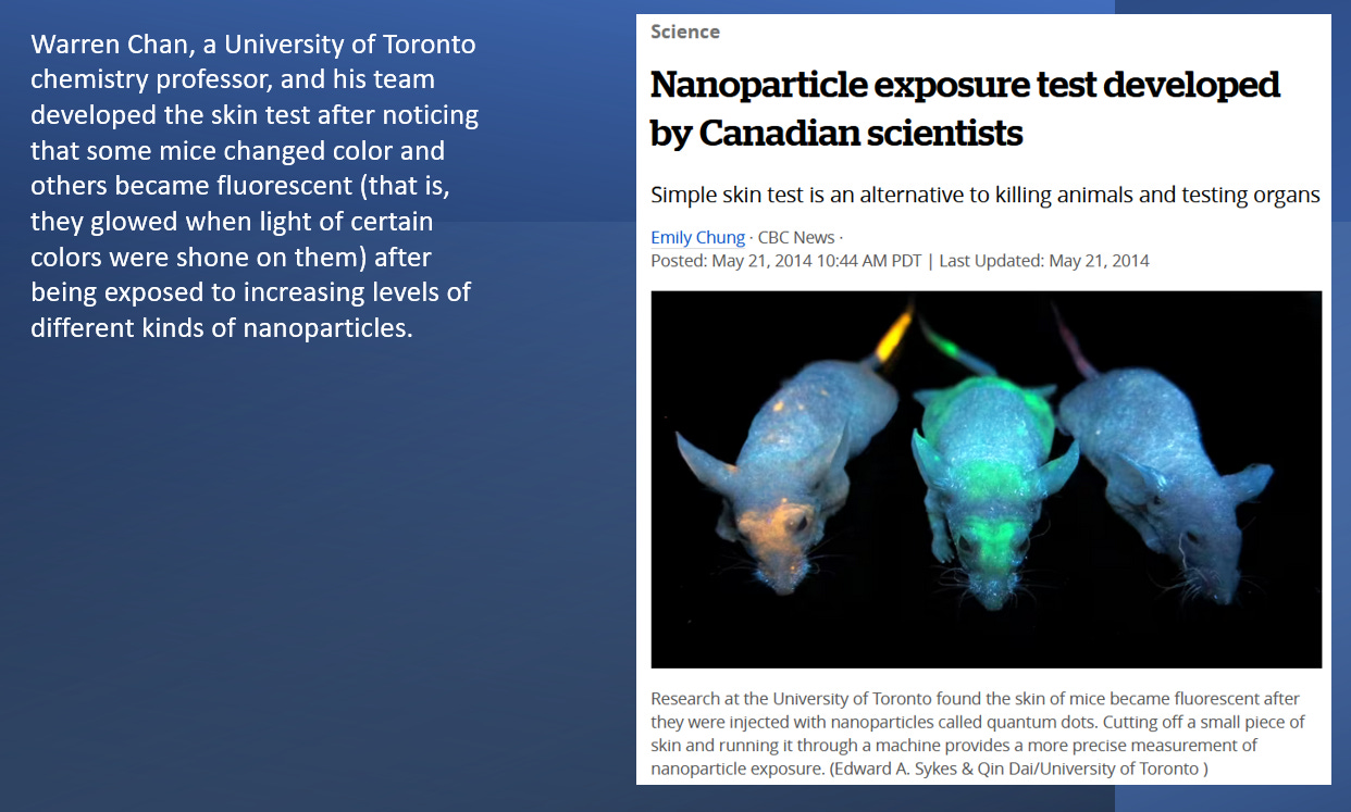
I showed the research where scientists in 2014 used light to determine nano particle exposure via fluorescent skin glow.
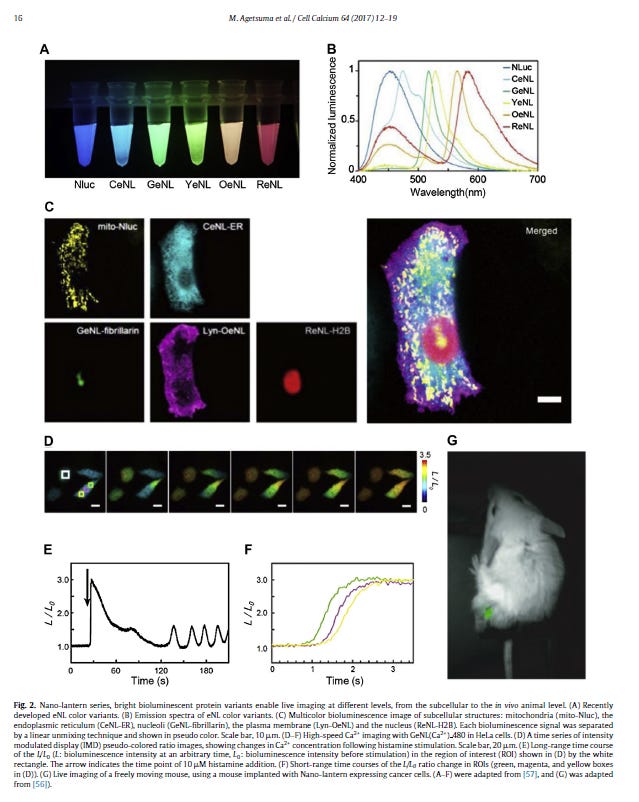
The neuroscience community is raving about the new green bio luminescent nano lanters that use an enzyme of a firefly ( note they are not saying LUCIFERASE - it might have dawned on them that the public is becoming sensitized to that word) that by the use of a “virus” is genetically encoded into the mice to express a fluorescent biosensor in the brain that allows the sensing and thereby imaging of oxygen.
The methods of nanolanterns using luciferase was described in Nature in 2013:
Takeharu Nagai of Osaka University and Hokkaido University and his colleagues now offer up a much brighter luminescent protein called Nano-lantern. This probe consists of an enhanced Renilla luciferase (RLuc) variant fused to the yellow fluorescent protein Venus. Venus serves as a BRET acceptor to RLuc's luminescent substrate coelenterazine to boost brightness. When expressed in HeLa cells, Nano-lantern allowed the researchers to record luminescence images that were nearly comparable to fluorescence images, even allowing the imaging of fine features such as microfilaments and microtubules .
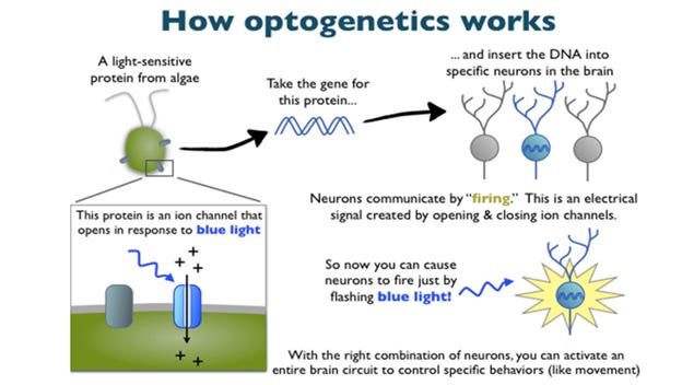
Here is the issue - if you turn on the light in the cells to sense different biochemical actions, you at the same time can CONTROL all aspects of the cells cycle via optogenetics.
The RLuc moiety of Nano-lantern contains an unstructured loop region into which functional sensor domains can be inserted. Nagai's team created luminescent sensors to observe Ca 2+ , cAMP and ATP dynamics in living cells. This allowed them to image situations in which it is not possible to use fluorescence, such as in combined Ca 2+ imaging with optogenetic control in neurons or in the detection of ATP in strongly autofluorescent plant chloroplasts.
In other words, if they want to study peoples brain by using fluorescent proteins, they can also control the brain. Of course, in the science literature everything is always written as if it is the best idea ever to evolve therapeutics. No mention ever that any of these technologies can be used in the most nefarious ways. This is where it is good to remember the military documents like Cyborg 2050 that describe that social acceptance of merging humans with machines will happen through healthcare, since everyone whats to cure Alzheimers or make a quadriplegic person mobile again.
Well, that comes with a pricetag, that might cost you not just your free will but possibly your soul.
Cyborg Soldier 2050: Human/Machine Fusion and the Implications for the Future of the DOD
The global healthcare market will fuel human/machine enhancement technologies primarily to augment the loss of functionality from injury or disease, and defense applications will likely not drive the market in its later stages. The BHPC study group anticipated that the gradual introduction of beneficial restorative cyborg technologies will, to an extent, acclimatize the population to their use.
A prime example of dual purpose applications were the C19 bioweapons, sold as safe and effective ways to protect you from a nonexistent virus while in actuality installing the whole body network interface for AI Cyborg remote control and upload to the metaverse.
You can read more about this here:
Optogenetic control of human neurons in organotypic brain cultures
Optogenetics is one of the most powerful tools in neuroscience, allowing for selective control of specific neuronal populations in the brain of experimental animals, including mammals. We report, for the first time, the application of optogenetic tools to human brain tissue providing a proof-of-concept for the use of optogenetics in neuromodulation of human cortical and hippocampal neurons as a possible tool to explore network mechanisms and develop future therapeutic strategies.
I have also written about optogenetics in depth in relation to the C19 bioweapons:
Here is the original article in Science behind a paywall:
Oxygen imaging of hypoxic pockets in the mouse cerebral cortex
Please note that nano lanterns are not new and they use Luciferase
Methods for monitoring signaling molecules in cellular compartments
This is what neuroscience news reported:
Summary:
A new study introduces a novel bioluminescence imaging technique for observing oxygen movement in mouse brains. This method, inspired by firefly proteins, reveals real-time, widespread patterns of oxygen distribution, offering insights into conditions like hypoxia caused by strokes or heart attacks.
It further explores how sedentary lifestyles could increase Alzheimer’s risk by detecting “hypoxic pockets” or areas of temporary oxygen deprivation. This research paves the way for better understanding diseases associated with brain hypoxia and testing therapeutic interventions.
Key Facts:
A novel bioluminescence imaging technique now allows scientists to observe oxygen movement in the brain, providing real-time, detailed views.
The method shows that areas of the brain can experience temporary oxygen deprivation, known as “hypoxic pockets,” which are more common in sedentary states and could be linked to an increased risk of Alzheimer’s disease.
This research, bridging work from the University of Rochester and the University of Copenhagen, can revolutionize our understanding of diseases associated with brain hypoxia and pave the way for new therapeutic interventions.
A new bioluminescence imaging technique, described today in the journal Science , has created highly detailed, and visually striking, images of the movement of oxygen in the brains of mice.
The method, which can be easily replicated by other labs, will enable researchers to more precisely study forms of hypoxia, such as the denial of oxygen to parts of the brain that occurs during a stroke or heart attack. It is already providing insight into why a sedentary lifestyle increases risk for diseases like Alzheimer’s.
“This research demonstrates that we can monitor changes in oxygen concentration continuously and in a wide area of the brain,” says Maiken Nedergaard, co-director of the Center for Translational Neuromedicine, which is based at both the University of Rochester and the University of Copenhagen.
“This provides us a with a more detailed picture of what is occurring in the brain in real time, allowing us to identify previously undetected areas of temporary hypoxia, which reflect changes in blood flow that can trigger neurological deficits” says Maiken Nedergaard.
Chemical cousin of Luciferase that is below.
Fireflies and serendipitous science
The new method employs luminescent proteins, chemical cousins of the bioluminescent proteins found in fireflies. These proteins, which have been used in cancer research, employ a virus that delivers instructions to cells to produce a luminescent protein in the form of an enzyme. When the enzyme encounters its substrate called furimazine, the chemical reaction generates light.
Like many important scientific discoveries, employing this process to image oxygen in the brain was stumbled upon by accident. Felix Beinlich, Assistant Professor at the Center for Translational Neuroscience at the University of Copenhagen, had originally intended to use luminescent proteins to measure calcium activity in the brain. It became clear there was an error in the protein production, causing a months-long research delay.
You can program the nanolanterns to measure anything, not just oxygen.
While Felix Beinlich waited for a new batch from the manufacturer, he decided to move forward with the experiments to test and optimize the monitoring systems. The virus was used to deliver enzyme-producing instructions to astrocytes, ubiquitous support cells in the brain that maintain the health and signaling functions of neurons, and the substrate was injected directly into the brain.
The recordings revealed activity, identified by a fluctuating intensity of bioluminescence, something that the researchers suspected, and would later confirm, reflected the presence and concentration of oxygen. “The chemical reaction in this instance was oxygen dependent, so when there is the enzyme, the substrate, and oxygen, the system starts to glow,” says Felix Beinlich.
Here is the catch - it does not just monitor oxygen tension but you can see that a motor action can immediately be monitored in the corresponding brain region:
Changes in light intensity also corresponded with sensory processing. For example, when the mice’s whiskers were stimulated with a puff of air, the researchers could see the corresponding sensory region of the brain light up.
“Hypoxic pockets” could point to Alzheimer’s risk
The brain cannot survive long without oxygen, a concept demonstrated by the neurological damage that quickly follows a stroke or heart attack. But what happens when small parts of the brain are denied oxygen for brief periods?
This question was not even being asked by researchers until the team in the Nedergaard lab began to look closely at the new recordings. While monitoring the mice, the researchers observed that specific tiny areas of the brain would intermittently go dark, sometimes for several seconds, meaning that the oxygen supply was cut off.
Oxygen is circulated throughout the brain via a vast network of arteries and smaller capillaries–or microvessels–which permeate brain tissue.
I have discussed the risk of having self assembly nanotechnology in your blood as these grow to a size hundreds and thousands of times larger than capillaries, hence would cause the very lack of oxygen described here.
Through a series of experiments, the researchers were able to determine that oxygen was being denied due to capillary stalling, which occurs when white blood cells temporarily block microvessels and prevent the passage of oxygen carrying red blood cells.
These areas, which the researchers named “hypoxic pockets,” were more prevalent in the brains of mice during a resting state, compared to when the animals were active. Capillary stalling is believed to increase with age and has been observed in models of Alzheimer’s disease.
Please note that long Covid, as I have long said is in the similar category as dementia which I have found on my functional EEG testing in my office and described a couple years ago, specifically targeting the hippocampus, the region of short term memory.
This was before I had a Darkfield microscope to see what is really in the blood, but the treatments used worked then and now. Unfortunately, the FDA pulled these life giving peptides off the market, so people would not have powerful reversal options of the depopulation efforts.
In the article you can see the extremely low brain voltage from “long Covid” aka exposure to self replicating nanotechnology in the blood.
COVID Spike Protein Causes Accelerated Aging
Continuing the article - discussing its future potential application in all aspects of heath:
“The door is open to study a range of diseases associated with hypoxia in the brain, including Alzheimer’s, vascular dementia, and long COVID, and how a sedentary lifestyle, aging, hypertension, and other factors contribute to these diseases,” says Maiken Nedergaard and adds:
“It also provides a tool to test different drugs and types of exercise that improve vascular health and slow down the road to dementia.”
Summary:
There is a lot of talk in the science community about bioluminescence and making everything on our planet glow, including humans. Remember that one sentence, that optogenetics is the most powerful tool - meaning the control of genes through light. You don’t want any novel light producing proteins in your brain to monitor anything.
Just like we do not want Quantum Dot microrobots swimming in our blood stream perform gene editing operations.
I would pay close attention to the FIREFLY commentary, aka Luciferase. The plan of the Luciferians is to tag everything with the signature of Lucifer and merge all life with Artificial Intelligence.
As long as there is life in us and the love of God vibrant, we should say NO to that.
Likes: 114 | Comments: 50 | Reposts: 21 | Share Options: Copy Link
Rachel - Apr 2
Rachel
Very simple test. Buy a UV light and check your water and pee in the dark. You'll be surprised. I recently was in London, the water was fluorescent and the pee even more. Less so in Geneva, Switzerland but still a bit, especially pee. If you excrete luminescent crap, it's because it's all over inside your body.
REPLY | 3 replies
Kellzilla - Apr 2
Kellzilla
Dr Ana Maria , i ran into this book today : The Indoctrinated Brain by Dr Michael Nehls. He outline how the spike blew up deep inside the brain , shrinking and damaged neuron repair centers. To make a short point he hinted Lithium orotate could repair and clear brain fog.
Is there something to it or they are normalizing taking low dose Lithium ?
Thnx in advance.,
REPLY | 10 replies
48 more comments...
anaunited anapost